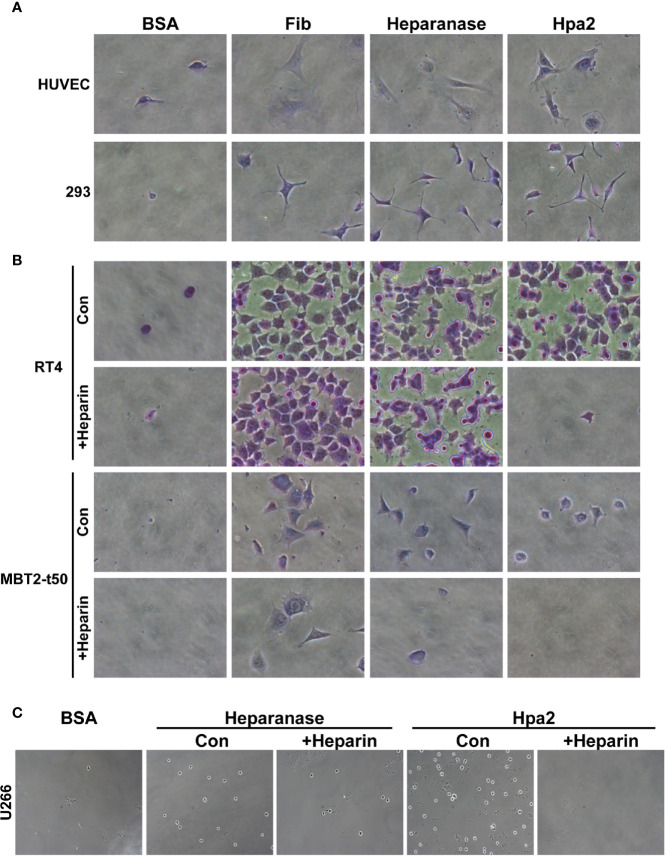
Figure 1.
Hpa2 promotes cell adhesion. Twenty-four–well plates were coated with 10 µg/ml of the indicated protein (18 h, 4°C), followed by blocking with BSA for 1 h at room temperature. The indicated cell type was then plated in growth medium and cell adhesion and spreading properties were observed. After 1 h, cells were washed, fixed, and photographed. Where indicated, heparin (50 µg/ml) was added together with the cells upon plating. (A) HUVEC (upper panels) and HEK 293 (lower panels). (B) RT4 human bladder (upper and second panels) and MBT2-t50 murine bladder carcinoma cells (third and fourth panels). (C) U266 myeloma cells. Shown are representative images at x50 (A, B), x25 (C) (original magnification).

