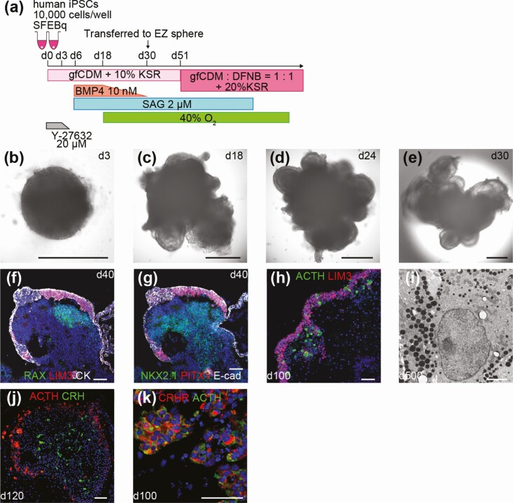
Figure 3.
In vitro hypothalamic-pituitary unit induced from human induced pluripotent stem cells (iPSCs). A, Modified condition for the human iPSC line. B-E, Self-formation of hypothalamic and pituitary tissues from human iPSCs. Double-layered structure was observed in aggregates. F, G, Both anterior pituitary and hypothalamic tissues were developed. LIM3 (f, red), PITX1 (g, red), RAX (f, green), NKX2.1 (g, green), pan-cytokeratin (f, white), E-cadherin (g, white). H, LHX3+ cells and ACTH+ cells were found to be abundant on day 100. LIM3 (red), ACTH (green). I, Electron micrograph of human iPSC-derived corticotrophs on day 500. J, ACTH+ cells and CRH+ cells coexisted in the same aggregates. ACTH (red), CRH (green). K, ACTH+ cells expressed CRHR. CRHR (red), ACTH (green). For all relevant panels, nuclear counterstaining was with DAPI (blue). Scale bars: 500µm (b-e), 50µm (f-h, j, k), 2µm (i).

