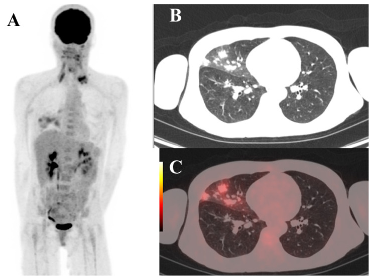
Figure 2.
Case 1. F18-FDG PET/CT scan images. MIP view (A) in a patient with nasopharyngeal cancer with pathologic F18-FDG uptake in left supraclavicular lymph nodes and in the right middle lung lobe. The CT axial view (B) revealed multiple lesions of “ground glass” in right middle lobe of the lung. The fused PET/CT axial section (C) shows high uptake of F18-FDG the right middle lobe lesions.

