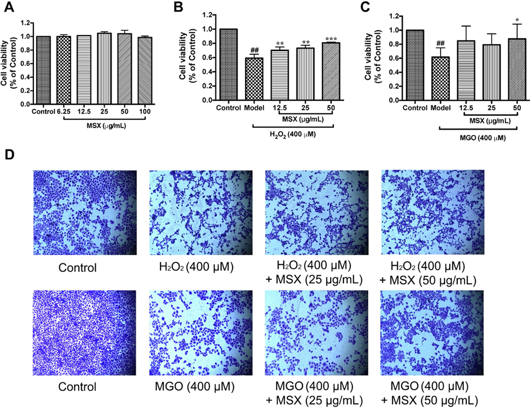
FIGURE 1.

Effects of MSX on the cell viability of H2O2 and MGO challenged HaCaT cells. MSX (6.25-100 μg/mL) were nontoxic to HaCaT cells, A. HaCaT cells were pretreated with MSX (12.5, 25, and 50 μg/mL) for 2 hours, then treated with H2O2 (400 μM; B), or MGO (400 μM, C). Representative images of cells stained with crystal violet reagent. HaCaT cells were pretreated with MSX and then exposed to H2O2 or MGO, D. ##Compared to control P < .01; *compared to model P < .05, **Compared to model P < .01, ***compared to model P < .001
