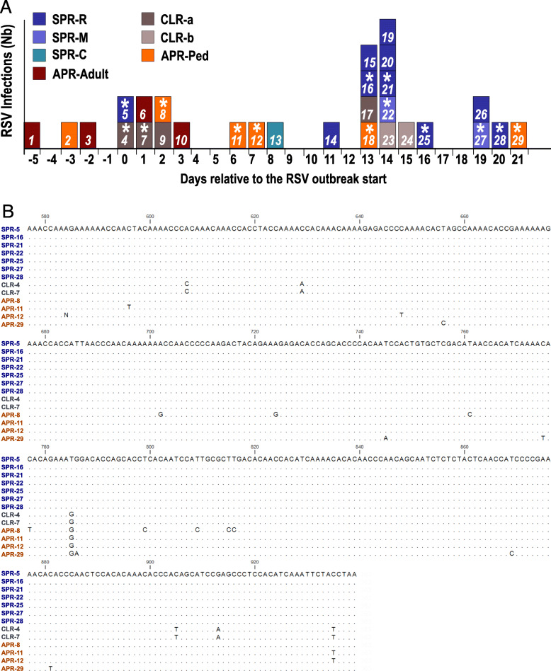
Fig. 1.
Chronology of the outbreak and molecular analysis. a: Distribution of confirmed RSV cases in the hospital group (GHUPIFO) in March in by onset of days relative to the outbreak start. The color identifies the ward involved. SPR-R, −M and -C are 3 adjacent wards in SPR building. Cases from SPR-R and -M (in blue) were considered as the “outbreak cases”. CLRa and b are 2 different wards from CLR building. The “*” symbol indicates successfully sequenced samples. b: Sequence alignment of the second variable region of the RSV G gene (nucleotide positions 577 to 939 relative to the start codon). Sequence of RSV-A (ON1) from patient 18 was not included in the alignment. Dots indicate nucleotide identities. RSV G gene nucleotide sequences were deposited in GenBank under accession numbers GenBank MT989450 to MT98945062

