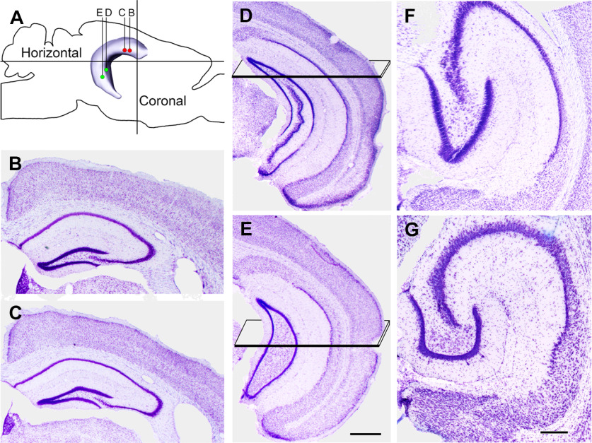
Figure 1.
Locations of the transfections and planes of section used for histologic analysis are illustrated diagrammatically and in Nissl-stained sections of the hippocampus. A, Transfections were made at two sites in the rostral (dorsal) and two sites in the caudal (ventral) DG. In double-labeled experiments, transfections for mCherry (red dots) were made rostrally, and those for eYFP (green dots) were made caudally. B–E, AP levels of the transfections are illustrated in coronal sections, progressing from the most rostral (B) to most caudal (E), and correspond to coordinates described in the text. Planes in D, E indicate the locations of the horizontal sections. F, G, For histologic analysis, the ventral hippocampus was sectioned in the horizontal plane, with the section in F corresponding to the region transected by the plane in D, and the section in G corresponding to the region transected by the plane in E. When sectioned in the horizontal plane, the rostral (anterior) region of the ventral DG has an elongated shape (F), and the more caudal region assumes a C-shape (G). The image of the hippocampus (A) and planes of section for the horizontal images (F, G) are based on data from the Allen Brain Atlas, Explorer 2. Scale bars: 500 µm (B–E) and 200 µm (F, G).

