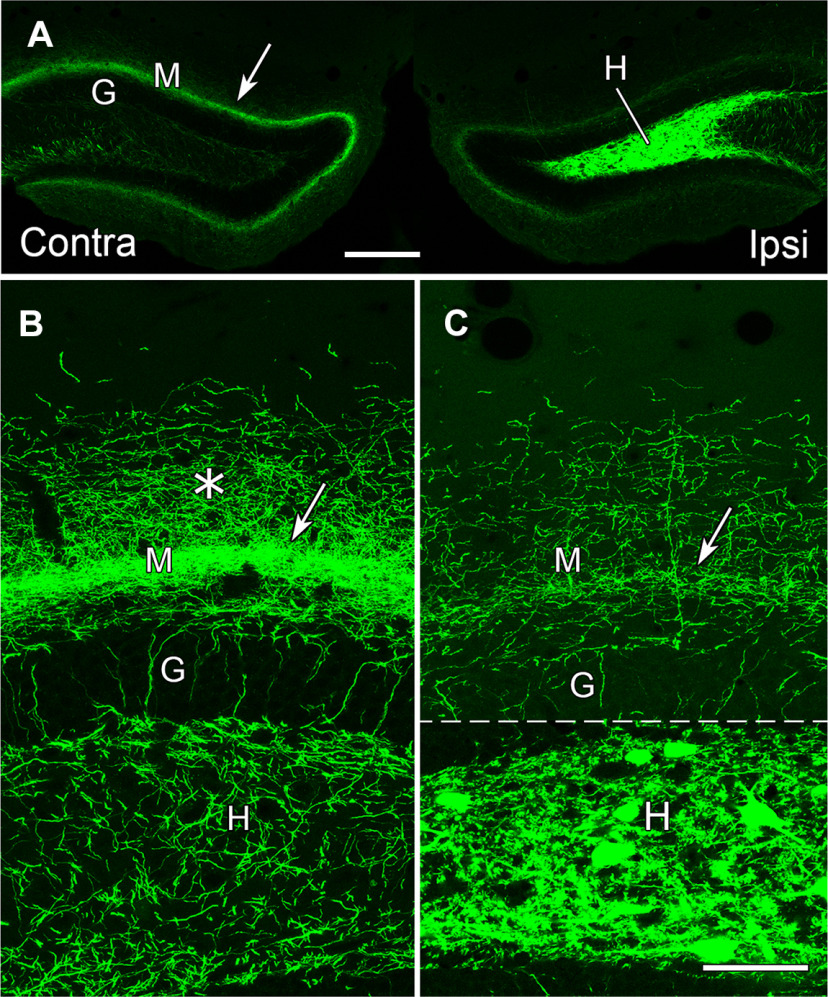
Figure 4.
eYFP-labeled MCs in the dorsal DG provide a direct commissural projection to the contralateral molecular layer and hilus at the level of the transfected neurons. A, Labeled neurons fill the dentate hilus (H) on the ipsilateral (transfected) side, and their axons form a distinct band (arrow) in the dentate molecular layer (M) of the contralateral side, adjacent to the unlabeled granule cell layer (G). B, In the contralateral DG, a narrow, dense band of labeled fibers (arrow) is evident at a slight distance from the granule cell layer (G) and a more diffuse plexus extends further into the molecular layer (*). Labeled fibers are also present in the contralateral hilus (H), and a few labeled fibers extend perpendicularly through the granule cell layer. C, On the ipsilateral side, numerous labeled cell bodies and processes are evident in the hilus. While labeled fibers are present in the inner molecular layer (arrow), their density is substantially lower than that on the contralateral side. This panel is a montage, with the regions above and below the dashed line imaged at different intensities, to avoid complete saturation of the strongly labeled cell bodies in the hilus when imaging the less intensely labeled plexus in the molecular layer. The region above the dashed line was imaged with the same parameters as those used for imaging the contralateral side, to allow comparison of the axonal plexuses. Scale bars: 200 µm (A) and 50 µm (B, C).

