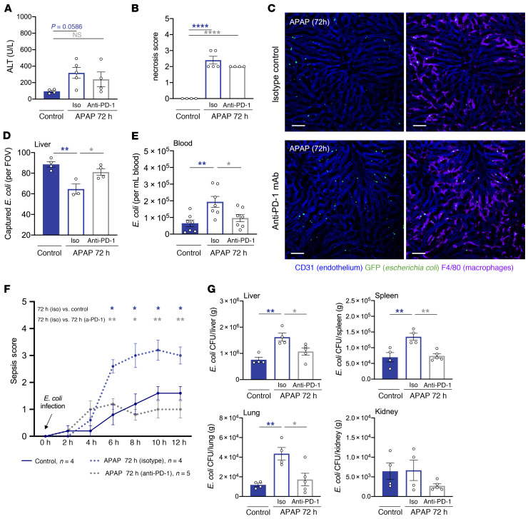
Figure 6. PD-1 blockade improves KC bacterial clearance and confers protection from sepsis in mice with acute liver injury.
Baseline (control) and APAP-treated (72 h) WT mice, dosed with isotype control (iso) or anti–PD-1 mAb 48 hours after APAP treatment, were intravenously challenged with E. coli. (A) Plasma alanine transaminase (ALT) levels (U/L) and (B) hepatic necrosis scores (n = 4–5 per group). (C) Representative intravital liver images from APAP-treated (72 h) mice (20 min after infection). Macrophages and endothelial cells were stained with fluorescently labeled anti-F4/80 (purple) and anti-CD31 (blue) antibodies, respectively; GFP+ E. coli (green). Scale bars: 50 μm. (D) Intravital liver imaging analysis showing the amount of macrophage-captured E. coli per FOV (n = 3–4 per group). (E) Number of free E. coli in blood measured by flow cytometry (n = 7 per group). (F) Mouse sepsis scores over time following E. coli infection (n = 4–5 per group). (G) CFU analysis showing bacterial burden in the liver, spleen, lungs, and kidneys 24 hours after infection (n = 4–5 per group). Results in A–G are for 2 independent experiments. Each symbol represents an individual mouse. Data are presented as the mean ± SEM. *P < 0.05, **P < 0.01, and ****P < 0.0001, by 1-way ANOVA (A–E and G) or 2-way, repeated-measures ANOVA (F).

