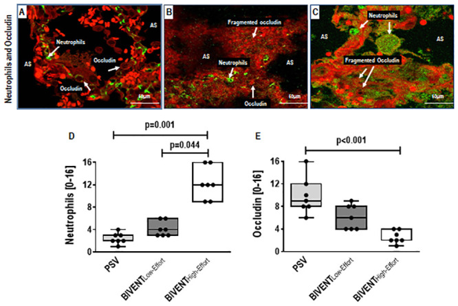
Fig 5. Double immunostaining of lung tissue for green-fluorescent neutrophils (GFN) and red-fluorescent occludin (RFO) from PSV, BIVENTLow-Effort, and BIVENTHigh-Effort animals, visualized under confocal microscopy.

Increased GFN cell counts (panel C; arrows) was seen in BIVENTHigh-Effort compared with PSV (panel A; arrows) and BIVENTLow-Effort (panel B; arrows) lungs. In contrast, increased RFO cell counts (panel A; arrows) were noted in PSV compared to both BIVENTLow-Effort and BIVENTHigh-Effort. Note the fragmented occludin in both BIVENT groups. AS: alveolar space. No difference in neutrophil counts was observed between PSV and BIVENTLow-Effort (p = 0.382). No differences in occludin were observed between PSV vs BIVENTLow-Effort and between BIVENTLow-Effort vs BIVENTHigh-Effort (p = 0.350 and p = 0.078, respectively). Box plots represent the median and interquartile range (panels D and E). Comparisons were done by the Kruskal–Wallis test followed by Dunn’s multiple comparisons (p<0.05).
