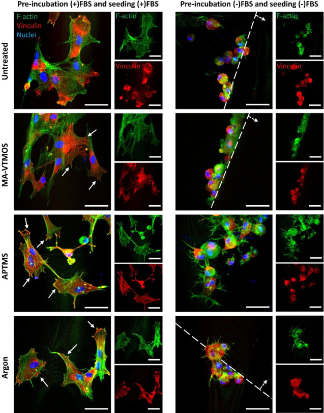
Figure 6.
HMSC morphology and focal adhesion formation after 4 h seeding on fresh plasma-treated scaffolds preincubated and seeded in (+)FBS or (−)FBS. Representative confocal microscopy images of hMSCs (F-actin, green; nuclei, blue; vinculin, red) on top of scaffold filaments 4 h post-seeding. Continuous arrows indicate focal adhesions. Dashed lines delimitate the scaffold filament, with dashed arrows indicating the filament’s surface. Scale bars 50 μm.

