
Figure 2.
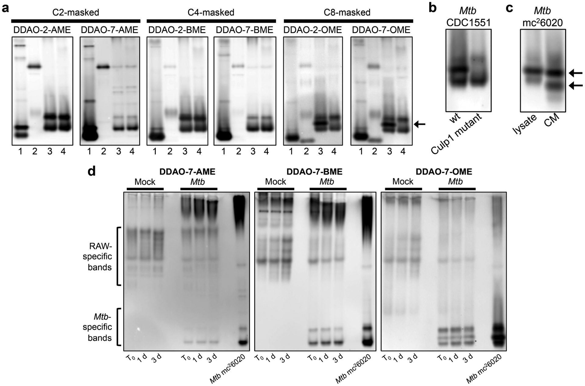
DDAO-derived fluorogenic probes reveal mycobacterial esterases and lipases in native PAGE-resolved lysates. (a) Lysates from (1) M. smegmatis, (2) M. marinum, (3) Mtb mc26020, and (4) M. bovis (BCG) were examined. The arrow highlights an Mtb-specific band revealed by the OME probes. (b) Lysates from Mtb CDC1551 wild type (wt) and a Culp1 transposon mutant confirmed that the Mtb-specific band corresponds to Culp1 activity. (c) Whole-cell lysate and conditioned medium (CM) from Mtb mc26020 both displayed Culp1 activity. Arrows indicate bands in the CM identified as Culp1 by mass spectrometry-based proteomics. (d) Mtb-infected RAW macrophages were collected and lysed immediately after the 4 h infection (T0), 1 day post-infection, or 3 days post-infection. Mock-infected macrophage lysates and Mtb mc26020 lysates were analyzed for comparison.