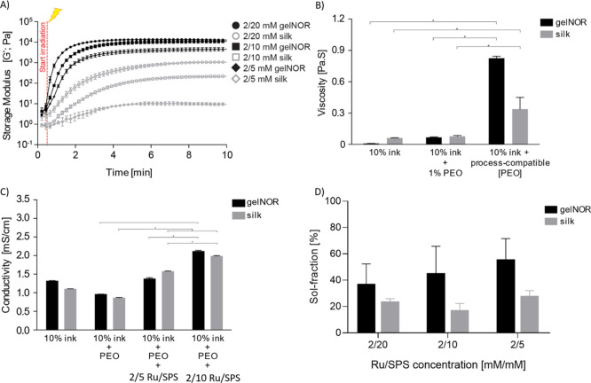
Figure 2.
Physical–chemical properties of gelnor and silk fibroin bioink systems. (A) In situ photorheometry showing the storage modulus (G′) of the bioinks as a function of time. Hydrogel samples were irradiated with visible light 30 s after the experiment started. (B) Viscosity changes as a function of the bioinks’ PEO concentration. (C) Electrical conductivity of hydrogels as a function of both PEO and photoinitiator concentration. (D) Sol fraction as a function of Ru/SPS photoinitiator concentration.* indicates significant difference.

