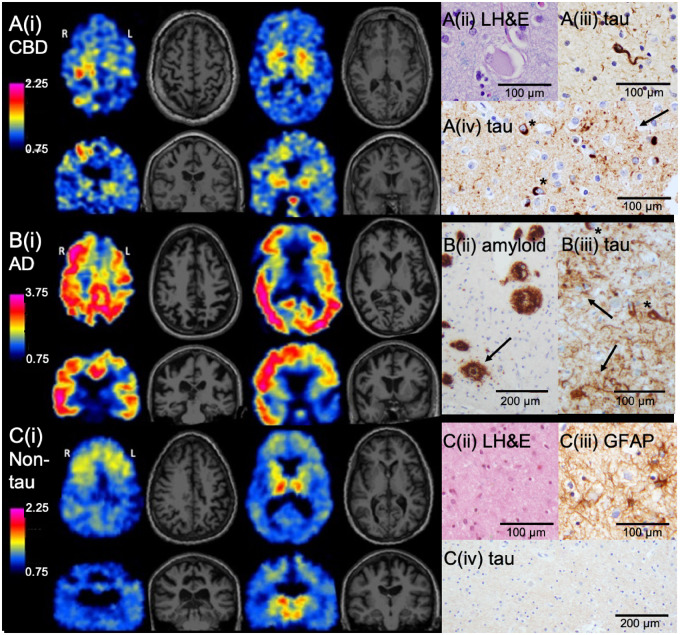
Figure 1.
Representative axial and coronal slices of 18F-AV-1451 PET and MRI and associated neuropathological findings in three cases of CBS. (A) CBS subject (aged 60 years) with left-predominant motor symptoms and neuropathologically confirmed CBD (Subject 2). [A(i)] 18F-AV-1451 retention was most notable in the right precentral gyrus and bilateral basal ganglia. Off-target binding was present in the pituitary. Neuropathological analyses confirmed CBD. [A(ii)] Rare ballooned cortical neurons were present in frontal cortex. [A(iii)] A moderate burden of tau-positive coiled bodies (oligodendrocyte cytoplasmic inclusions) was present in underlying white matter. [A(iv)] The burden of cortical tau pathology was greatest in the right precentral gyrus (shown), compared with the parietal, occipital, and temporal lobes. Immunohistochemistry with anti-tau antibody demonstrated numerous astrocytic plaques (arrow) and intraneuronal inclusions (asterisks). (B) CBS subject (aged 52 years) with left predominant motor symptoms and neuropathologically confirmed Alzheimer’s disease (AD) (Subject 8). [B(i)] Widespread but asymmetric cortical 18F-AV-1451 retention was present. Note the modified colour scale, with higher upper value, to capture the high levels of tracer retention present in this Alzheimer’s disease case. [B(ii)] Immunohistochemistry with anti-amyloid-β antibody demonstrated a heavy burden of dense-cored (arrow) and diffuse plaques. Parietal cortex is shown. [B(iii)] Anti-tau antibody in parietal cortex showed numerous neuritic plaques (arrows) and neurofibrillary tangles (asterisks). (C) CBS subject (aged 59 years) with left predominant motor symptoms without evidence for tau pathology on neuropathological assessment (Subject 11). [C(i)] 18F-AV-1451 retention was evident in the bilateral thalami and the bilateral substantia nigra, and to a lesser extent symmetrically in the frontal subcortical and perithalamic white matter and the basal ganglia. [C(ii)] Neuropathology demonstrated prominent gliosis with neuronal dropout in the thalamus, including in the dorsomedian nucleus (shown). [C(iii)] Immunohistochemistry with anti-GFAP antibody showed numerous reactive astrocytes in the thalamus. [C(iv)] Immunohistochemistry with anti-tau antibody in the thalamus (shown) and other regions was negative for abnormal tau accumulation. 18F-AV-1451 SUVR scales are shown for each subject. GFAP = glial fibrillary acidic protein; LH&E = Luxol fast blue/haematoxylin and eosin.

