. 2021 Jan 3;35(1):249–257. doi: 10.21873/invivo.12253

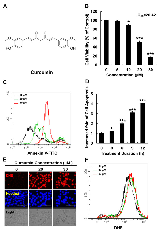
Figure 1. Curcumin increased cellular ROS levels and induced apoptosis in HepG2 liver cancer cells. (A) The chemical structural of curcumin. (B) Cell viability was analyzed using MTT assay, in HepG2 cells treated with curcumin (0, 5, 10, 20, or 30 μM) for 12 h. (C) Cellular apoptosis was measured using flow cytometry in HepG2 cells treated with curcumin (0, 20, or 30 μM) for 12 h. Black line: without curcumin; green line: 20 μM curcumin; red line: 30 μM curcumin. (D) Cellular apoptosis was measured by flow cytometry in HepG2 cells treated with curcumin at different times (0, 3, 6, or 12 h). The fold increase in apoptotic cells is presented as the mean±SEM (*p<0.05, **p<0.01). (E) Cellular ROS levels were detected by fluorescence microscopy in DHE (red)- and Hoechst (blue)-stained HepG2 cells treated with curcumin (0, 20, or 30 μM) for 12 h (scale bar=100 μm). (F) Cellular ROS levels were detected by fluorescence microscopy in DHE-stained HepG2 cells treated with curcumin (0, 20, or 30 μM) for 12 h.

Figure 1