
Figure 3. mtCU inhibition by MCU-i11 in WT, MICU1KO and MCUKO cell lines.
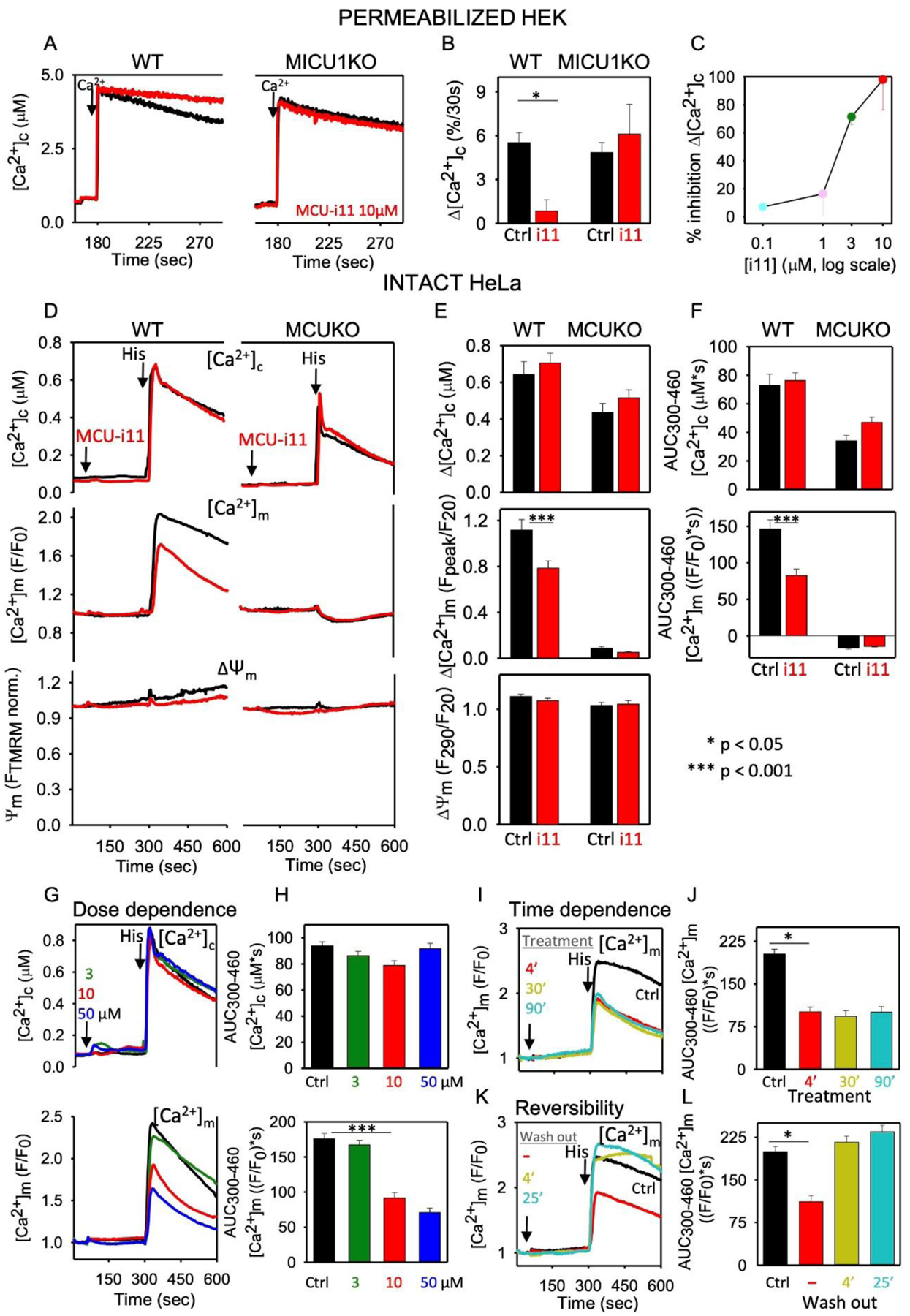
A Clearance of a Ca2+ bolus (10μM CaCl2) was monitored in saponin-permeabilized WT and MICU1KO HEK cells in the presence of 2μM thapsigargin and 10μM CGP37157 to inhibit ER Ca2+ uptake and NCLX, respectively as described in [54]. Clearance of Ca2+ was completely prevented by RuRed (100%, n=3), confirming that clearance was due to mitochondrial Ca2+ uptake via the mtCU. Representative traces show the inhibition of mitochondrial Ca2+ uptake by 10μM MCU-i11 in WT, and the loss of inhibition in MICU1KO. B Percentage of Ca2+ uptake (30s) inhibition by MCU-i11 (i11). C Dose response curve of MCU-i11’s inhibition of mitochondrial Ca2+ uptake (n=6). D [Ca2+]c (fura2), [Ca2+]m (mtCepia) and ΔΨm (TMRM) were simultaneously monitored in intact wild type (WT) and MCUKO HeLa cells treated sequentially with 10 μM MCU-i11/DMSO and 100μM histamine [45]. Traces represent the means of cells from 6–8 measurements of 2 experimental days. E Baseline subtracted peak amplitude of [Ca2+]c and [Ca2+]m, and the ΔΨm measured at the time of the peak normalized to the prestimulation value. F AUC for [Ca2+]c and [Ca2+]m. GH MCU-i11 (3, 10, or 50 μM) was added to WT HeLa cells before stimulation with histamine. Mean [Ca2+]c and [Ca2+]m traces in G and AUC in H. IJ WT HeLa were treated with 10μM MCU-i11 for 4, 3, or 90min, before histamine addition. Mean [Ca2+]m traces in I and AUC in J for the results of quadruplicate measurements for each condition. KL Intact WT HeLa cells were pretreated with 10μM MCU-i11 for different intervals (4, 30, 90min) and then it was washed out, or not, 4 or 25min before stimulation with histamine. Mean [Ca2+]m traces in K and AUC in L. Every single cell fluorescence imaging experiment was reproduced with ≥3 different cell cultures in quadruplicate measurements each day. Results are expressed as mean ± SEM (*p<0.05, ***p<0.001).