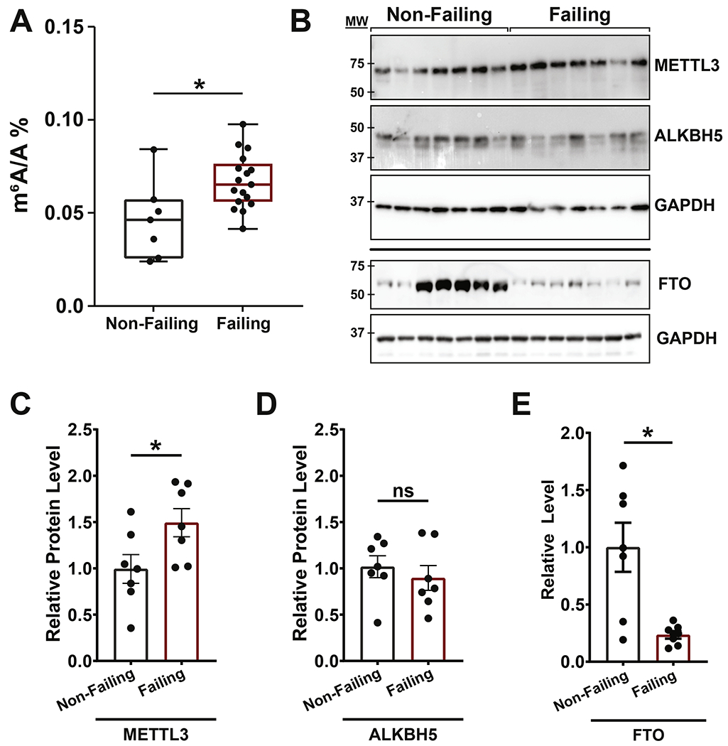
Figure 1. m6A and associated catalyzing enzyme are enriched in failing human heart tissue.

(A) RNA isolated from non-failing and failing human heart samples analyzed by UHPLC-MS/MS for global levels of methyl-6-adenosine (m6A) in relation to unmodified adenosine (A) showed a significant increase in total m6A in failing hearts (non-failing: n = 7; failing: n =17). (B) Western blots on non-failing and failing heart protein lysates measuring expression of METTL3 (m6A methylase), ALKBH5 and FTO (m6A demethylases). GAPDH and ponceau staining are shown as loading controls. (C-D) Quantification of METTL3 (C), ALKBH5 (D) and FTO (E) protein amount using GAPDH as control showing increased levels of METTL3 and decreased levels of FTO in failing hearts while ALKBH5 levels were stable (non-failing and failing: n = 7). Dots on graphs indicate individual data point from biological replicates, ns = nonsignificant, * = p < 0.05. Statistics carried out using two tailed t-test.
