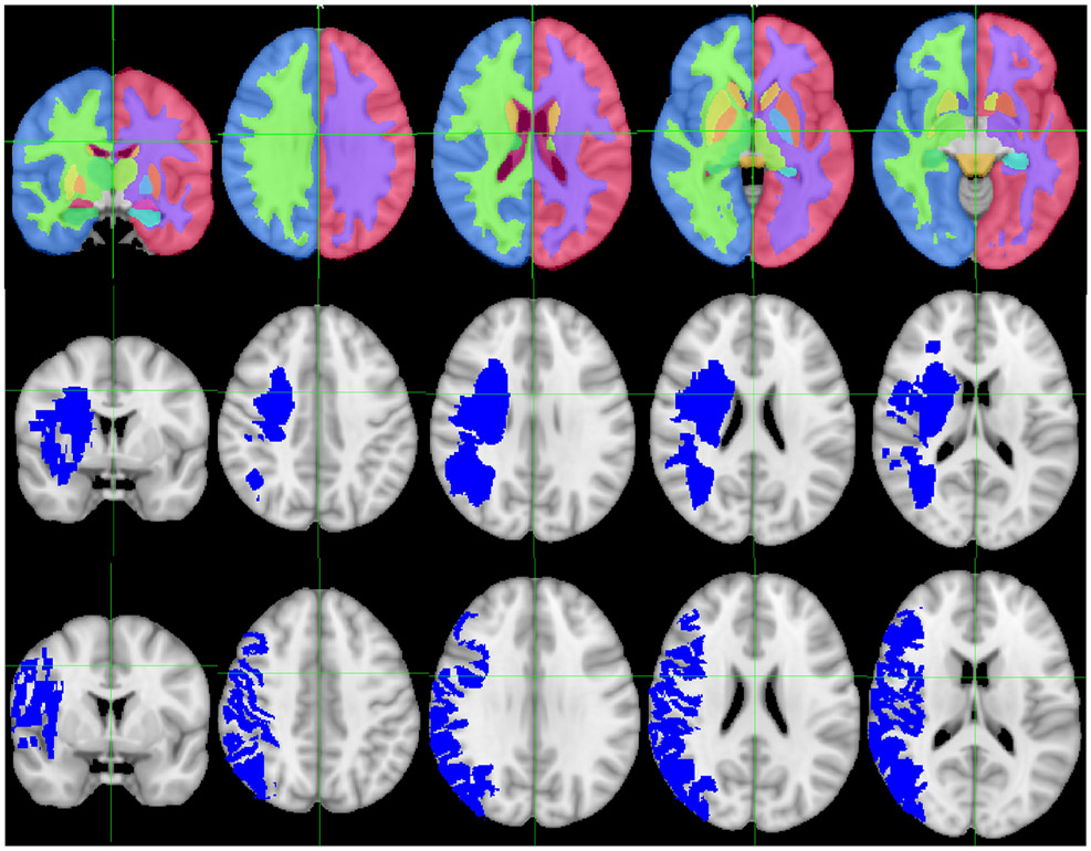
Figure 1.
Region-specific infarct volumes. Top row: Sections through the Harvard-Oxford subcortical atlas, demonstrating white matter, cortex, and basal ganglia regions, overlaid on the MNI 152 standard brain. Middle row: Sections through an example of an infarct affecting primarily white matter overlaid on the MNI 152 standard brain. Bottom row: Sections through an example of an infarct affecting primarily cortex overlaid on the MNI 152 standard brain.

