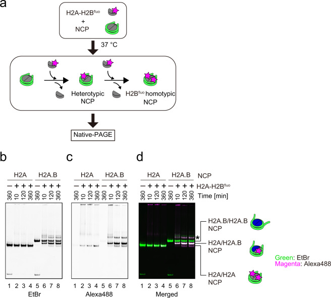
Fig. 2. Histone exchange activity of the H2A.B NCP.
a Schematic representation of the histone exchange assay. The NCP was incubated with the H2A-H2B dimer, in which the H2B protein was conjugated with the Alexa Fluor 488 fluorescent dye (H2A-H2Bfluo dimer). The samples were analyzed by native-PAGE. b–d Representative gel images of the histone exchange assay. The H2A NCP (1 µM) was incubated with the H2A-H2Bfluo dimer (0 µM: lane 1; 2 µM: lanes 2, 3, and 4) for 10 (lane 2), 120 (lane 3), and 360 (lanes 1 and 4) minutes. The H2A.B NCP (1 µM) was incubated with the H2A-H2Bfluo dimer (0 µM: lane 5; 2 µM: lanes 6, 7, and 8) for 10 (lane 6), 120 (lane 7), and 360 (lanes 5 and 8) minutes. After the incubation, the samples were analyzed by native-PAGE. The gels were visualized by ethidium bromide staining, or through the Alexa488 conjugated with the H2A-H2B dimer. b Ethidium bromide; c Alexa488 (H2A-H2Bfluo); d Overlay of ethidium bromide (green) and Alexa-488 (magenta). The asterisk indicates the NCP-histone complexes. Reproducibility is confirmed by three independent experiments, and the results are presented in Supplementary Fig. 3. The uncropped gel images are shown in Supplementary Fig. 9.

