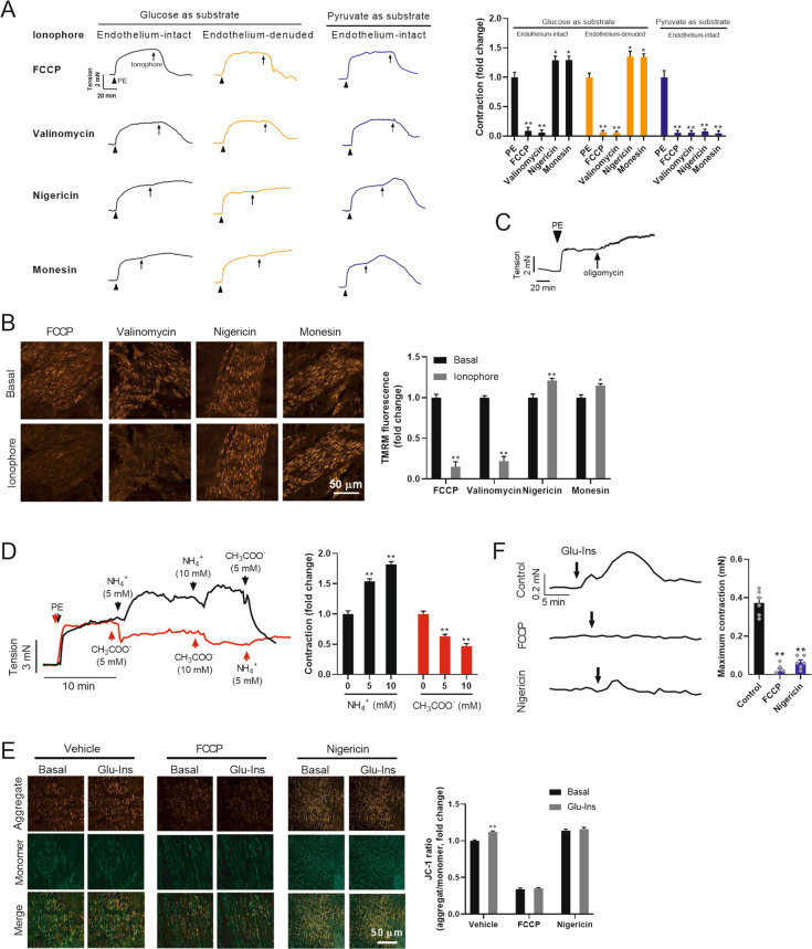
Fig. 3. ΔΨm positively regulates VSMC contraction.
A The effects of four ionophores on vascular constriction in isolated thoracic aortas with intact endothelium or with denuded endothelium. The aortas were pretreated with PE. Glucose (5 mM) or pyruvate (5 mM) was supplied as the only metabolite available in the incubation medium. Typical traces of vascular tension were shown in the left, and the quantified results were shown in right. The arrow indicates the time point for treatment with ionophore. n = 6. B The effects of four ionophores on ΔΨm in VSMCs of isolated aortas. Typical images of ΔΨm in VSMCs in blood vessels were shown in the left, and the quantified results were shown in right. ΔΨm was detected by TMRM. n = 6. C Oligomycin induced vascular constriction in isolated thoracic aortas. D The effects of NH4+ and CH3COO- on vascular tone in isolated aortas. Typical traces of vascular tension were shown in the left, and the quantified results were shown in right. n = 6. E Glu-Ins-induced mitochondrial hyperpolarization was blocked by preincubation of isolated aortas with FCCP or nigericin. ΔΨm was detected by JC-1. Typical images of ΔΨm in VSMCs in blood vessels were shown in the left, and the quantified results were shown in right. F Preincubation of isolated aortas with FCCP or nigericin inhibited Glu-Ins-induced vascular constriction. Typical traces of vascular tension were shown in the left, and the quantified results were shown in right. n = 6. Error bars represent SEM. *P < 0.05. **P < 0.01.

