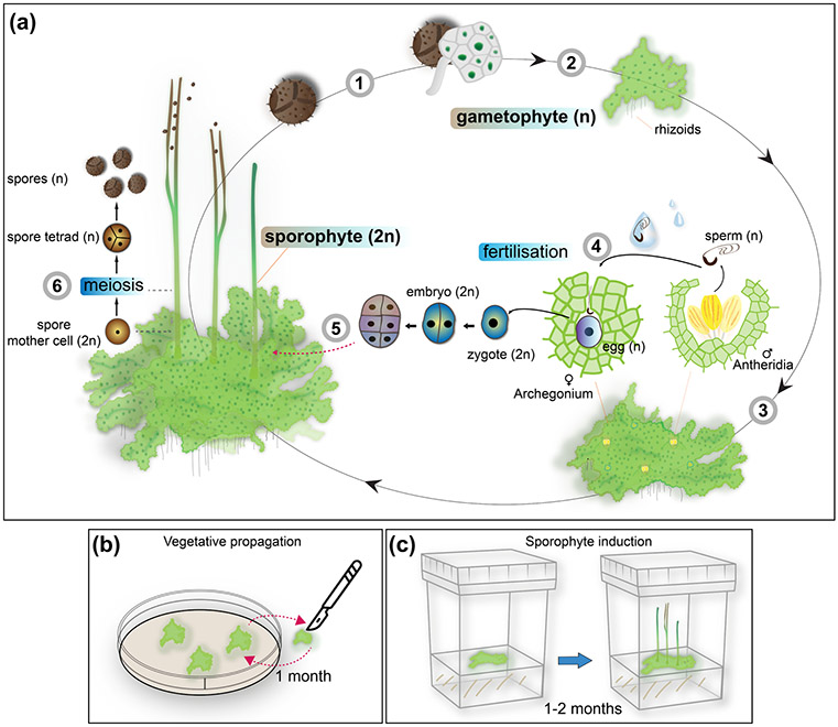
Fig. 1: Life and laboratory cycle of the hornwort A. agrestis.
(a) A. agrestis has two life cycle phases. A dominant haploid phase called gametophyte and a diploid phase called sporophyte. The life cycle of A. agrestis starts with the germination of the haploid spores (1) which develop into an irregularly shaped thallus (2). A. agrestis is monoecious, with both male and female reproductive organs present on the same individual. Male (antheridia) and female (archegonia) reproductive organs are embedded in the thallus and mitotically produce sperm and egg, respectively (3). Biflagellated motile sperm cells swim in water to the archegonium where the egg is fertilised (4). The resulting diploid zygote divides first by a longitudinal division and subsequent divisions to form the embryo, which is initially composed of three tiers. The bottom tier produces the foot. The middle tier gives rise to the basal meristem and the top tier forms the tip of the sporophyte (5). The sporophyte develops within the gametophyte and is nourished through the placenta, the junction between foot and gametophyte cells. Meiosis and sporogenesis occur progressively from the base of the sporophyte upwardly, leading to spore formation: sporogenous tissue at the base of the sporophyte produces spore mother cells that, via meiosis, produce spore tetrads and spores that are released at the tip where the sporophyte separates into two valves. (6). n: haploid, 2n: diploid.
(b - c) Laboratory cycle of A. agrestis: (b) Plants can be easily propagated in axenic culture by transferring small thallus fragments (typically 1 mm x 1 mm) onto plates with fresh growth media using sterile scalpels. (c) In laboratory conditions A. agrestis sporophyte induction can be achieved in 1-2 months under axenic conditions using a small thallus fragment as starting material.

