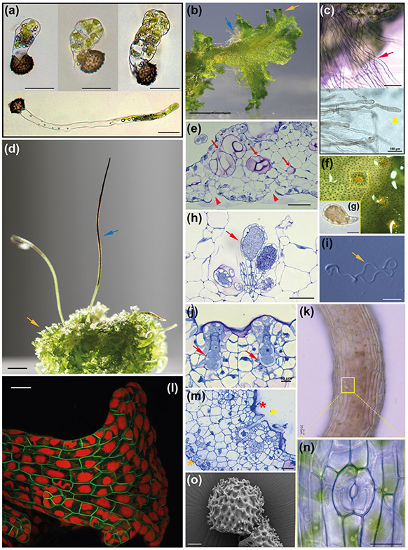
Fig. 2: Key morphological features of A. agrestis.
(a) Light micrograph (LM) of germinating spores. Upper three images, successive stages in globose sporeling production. Lowermost: under low light conditions spore germination involves a germ tube, a long single-celled filament that develops a terminate globose sporeling. Scale bars: 50 μm. (b) Surface view of the irregularly shaped thallus. Blue arrow: rhizoids. Orange arrow: wavy thallus edge. Scale bar: 2.0 mm. (c) Top: LM of single-celled rhizoids on the ventral thallus (red arrow). Scale bar: 150 μm. Bottom: LM of single-celled rhizoid tips (yellow arrowhead) Scale bar: 100 μm (d) Sporophytes (blue arrow) growing on the gametophyte (yellow arrow). Scale bar: 3.0 mm. (e) LM of longitudinal section of thallus with mucilage canals (red arrows). Mucilage clefts on the ventral side indicated with red arrowheads. Scale bar: 50 μm. (h) LM of antheridia (red arrow) in an antheridial chamber in longitudinal section. Scale bar: 50 μm. (f) Surface view of antheridial chamber with yellow antheridia embedded in the dorsal thallus. Scale bar: 10 μm (h). (g) Antheridium removed from chamber in (f) showing antheridial body with sperm cells inside and stalk to the lower right. LM of two archegonia embedded in the dorsal thallus in longitudinal section showing from the base up: egg cell, ventral canal cell, neck cells and cover cells. Scale bar: 10 μm. (i) LM of biflagellate sperm. Coiled cell body is on the left and the flagella are on the right (yellow arrow). Scale bar: 5.0 μm. (m) LM longitudinal section of thallus with an open archegonium containing only the egg cell near the apical notch. Dorsal side (red asterisk) and ventral side (orange asterisk). Scale bar: 25 μm. (k) Sporophyte with stomata. Scale bar: 20 μm. (l) Confocal fluorescence microscopy image of transgenic gametophyte showing single plastids in each cell. Green: green fluorescent protein localised in the plasma membrane expressed under the CaMV 35S promoter. Red: chlorophyll autofluorescence. Scale bar 50 μm. (m) (n) Higher magnification LM of a stoma with two guard cells surrounding a pore. Scale bar: 10 μm. (o) Scanning electron micrograph (SEM) of distal side of a spinose spore. Scale bar: 10 μm.

