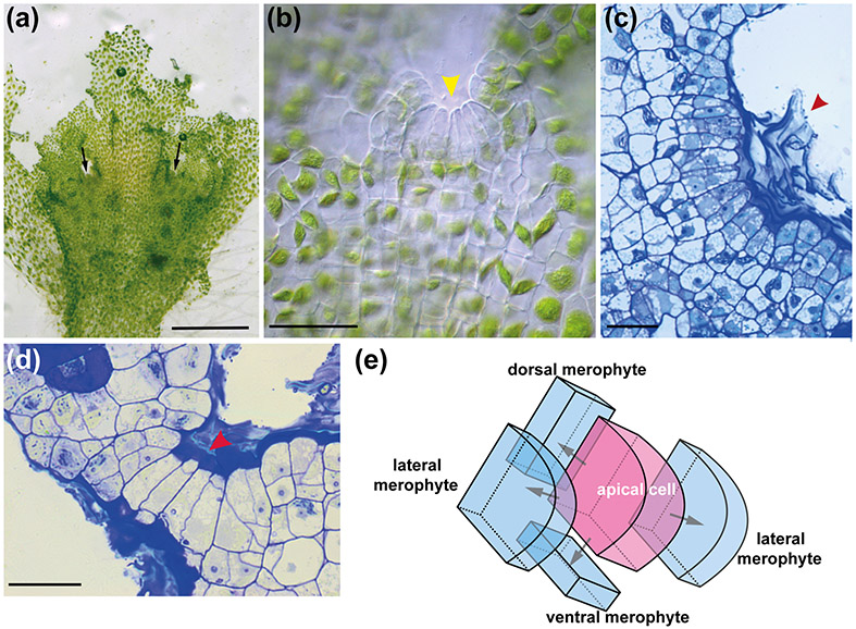
Fig. 4 : A. agrestis gametophyte apical growth.
(a) Surface view of A. agrestis gametophyte. Arrows indicate apical notches. Scale bar: 1.0 mm (b) LM surface view of apical notch (yellow arrowhead) showing row of apical cell and immediate derivatives and single chloroplasts in older cells. Scale bar: 50 μm (c) LM surface section of apical notch covered by mucilage (arrowhead). Scale bar: 50 μm. (d) LM transverse section of thallus showing four rectangular cells that include the apical cell (red arrowhead) and three immediate derivatives in a growing notch covered by mucilage. The more abundant cells on either side are from divisions in the lateral derivatives. Scale bar: 50 μm. (e) Schematic representation of gametophyte apical cell (pink) with four cutting faces and four derivatives (blue).

