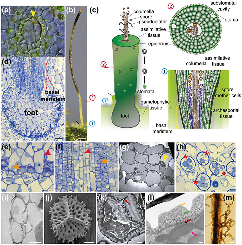
Fig. 6: A. agrestis embryo and sporophyte.
(a) Differential interference contrast image of embryo with first longitudinal division (yellow arrowhead). Scale bar 10 μm. (b) Sporophyte. Spores mature progressively from the bottom to the top of the sporophyte. Involucre indicated with black arrow. Scale bar: 0.5 mm. (c) Schematic representation of sporophyte. The sporophyte has a foot, a basal meristem, the columella, a spore layer with pseudoelaters (for spore dispersal), a multicellular assimilative layer, and stomata. Stomata successive developmental stages are shown next to the sporophyte. Opening of the pore occurs near the base of the sporophyte then guard cells wall thickens, guard cells collapse toward the upper part of the sporophyte, allowing dehydration and dehiscence into two valves. Involucre indicated with black arrow. Numbered circles indicate the relative position on the sporophyte that corresponds to the cartoons on the right of panel: 1. Schematic representation of longitudinal section directly above the foot showing the basal meristem and differentiating sporogenous tissue. 2. Schematic representation of transverse section showing from centre to outside, columella, spores, pseudoelaters, assimilative tissue, epidermis and stomata with substomatal cavities. (d) LM longitudinal section of bulbous foot and basal meristem surrounded by the involucre. The placenta consists of elongated haustorial foot cells adjacent to small gametophyte cells. Scale bar: 50 μm. (e) Enlargement of placental cells in (d) showing smooth walled haustorial cells (red arrowhead) intermixed with gametophyte cells (orange arrowhead) with conspicuous cell wall ingrowths (orange arrowhead) Scale bar: 10 μm (f) LM longitudinal section of archesporial tissue (orange arrowhead) surrounding columella (red arrowhead) directly above the basal meristem. Scale bar: 20 μm (g) Transmission electron microscopy (TEM) image of columella in cross section showing 4 by 4 arrangement of the 16 living cells that contain dense cytosol and chloroplast (yellow arrowhead). Scale bar: 4.0 μm (h) LM section showing spore mother cells (red asterisks) with three of four large starch-filled plastids in four poles and central nucleus preparing for meiosis. Scale bar: 20 μm (i) TEM of dying and collapsing stoma (brown in (c)) showing thickened walls, inner and outer ledges of guard cells, and substomatal cavity. This section is on the polar end of the guard cells, away from the pore. Scale bar: 5 μm (j) SEM of a proximal surface of spore with a defined trilete mark. Scale bar: 10 μm. (k) TEM of spore in a tetrad still surrounded by the spore mother cell wall showing three-layered wall with ornamentation. The aperture on the proximal wall where spores in a tetrad meet each other has a thick intine and includes the trilete mark (red arrow). Scale bar 4.0 μm. (l) TEM of mature spore wall composed of outer exine (orange arrow), thick inner exine with compressed globular sporopollenin (green arrow) and thin intine that is much like a primary cell wall (red arrow). Protein bodies fill the spore (pink arrow). Scale bar 0.5 μm. (m) LM of dissected columella with elongated multicellular pseudoelaters attached. Scale bars: 25 μm.

