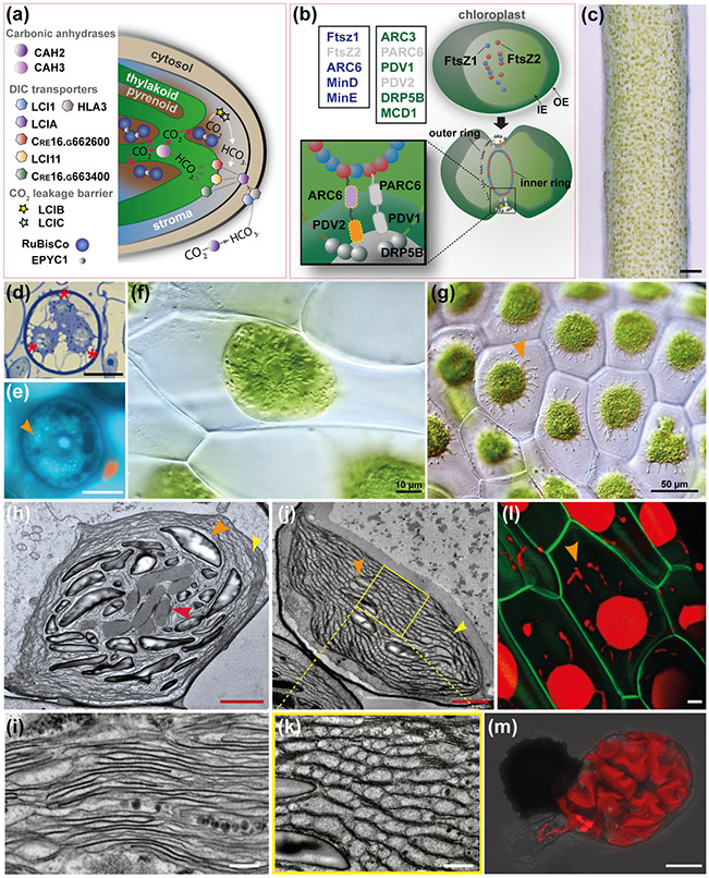
Fig. 7: A. agrestis chloroplast.
(a) Schematic representation of C. reinhardtii CCM model. The carbonic anhydrase (CAH2) converts CO2 into HCO3− (dicarboxylate (DIC)) in the periplasmic space. DIC is pumped across membranes via DIC transporters localised in the plasma membrane (low-CO2 inducible 1 (LCI1) and high light activated 3 (HLA3)), the chloroplast envelope (low-CO2 inducible A (LCIA)) and the thylakoid membrane (low-CO2 inducible 11 (LCI11), Cre16.g662600 and Cre16.g663400). The carbonic anhydrase 3 (CAH3) in the thylakoid lumen converts HCO3− into CO2. supplied to RuBiSCo. EPYC1 is acting as glue between RuBiSCo units in the pyrenoid. The low-CO2 inducible B and C (LCIB/C) proteins are thought to form a molecular “ring” around the pyrenoid that acts as a barrier to CO2 leakage transferring CO2 back to the thylakoid via the DIC pumps. (b) Top left boxes: Genes of prokaryotic origin (in blue) and genes of land plant origin (in green) involved in chloroplast division in A. thaliana. Genes absent in A. agrestis genome are in grey. Top right and bottom: Schematic representation of the key elements of chloroplast division machinery in A. thaliana (Chen et al., 2018). FtsZ1 and FtsZ2 self-assemble and then recruit ARC6, PARC6, PDV1, PDV2 and DRP5B forming a ring around the chloroplast that mediates its division. IE: inner envelope, OE: outer envelope. (c) Surface view of sporophyte showing chloroplasts. Scale bar: 100 μm. (d) LM section of spore mother cell with plastids (three out of four visible) at poles indicated with asterisks. Scale bar: 15 μm (e) LM of spore mother cell stained with DAPI showing DNA in central nucleus and plastids (orange arrowhead). Scale bar: 15 μm (f) Cells of young gametophyte tissue with a single chloroplast and central pyrenoid surrounded by starch grains. Scale bar: 10 μm (g) Cells of mature gametophyte tissue with single chloroplasts that have several protrusions that are likely stromules (orange arrowhead). Scale bar: 50 μm. (h-k) TEMs of chloroplasts of A. agrestis. (h) Chloroplast in an assimilative cell of sporophyte near intercellular space, with pyrenoid (red arrowhead) traversed by thylakoids and small grana (orange arrowhead). Starch granules indicated by yellow arrowhead and thylakoids enlarged in (i) at yellow arrowhead. Scale bar: 2.0 μm. (i) Details of thylakoids and grana showing absence of end membranes. Scale bar: 0.5 μm. (j&k) Sporophyte chloroplast in an assimilative cell near intercellular space traversed by channel thylakoids and grana stacks (yellow arrowhead). Starch grains indicated by orange arrowhead. Pyrenoid is not in this non-median section. Scale bar: 500 nm. (k) Higher magnification (scale bar: 0.5 μm) of region indicated in (j) showing channel thylakoids (red arrowhead). Scale bar: 2.0 μm. (l) Confocal microscopy image of A. agrestis transgenic gametophyte. Green: green fluorescent protein localised in the plasma membrane expressed under the CaMV 35S promoter. Red: chlorophyll autofluorescence. Orange arrowhead indicates chloroplast protrusions likely to be stromules. Scale bar: 10 μm (m) Confocal microscopy image of a germinating spore highlighting the wavy 3D structure of the chloroplast. Red: chlorophyll autofluorescence. Scale bar: 20 μm.

