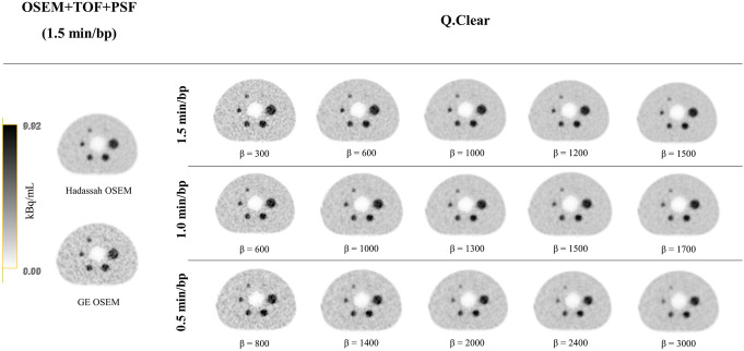
Fig. 2.
Central slice of the NEMA IEC image quality body phantom reconstructed with the Hadassah and GE OSEM reconstruction algorithms (1.5 min/bp) and with Q.Clear for different values of β and acquisition time per bed position (1.5, 1.0 and 0.5 min/bp). The phantom background region and spheres contained a 68Ga activity concentration of 2.48 kBq/mL and 9.92 kBq/mL (4:1 sphere-to-background ratio), respectively, at the time of acquisition. The gray scale represents the activity concentration in kBq/mL for all phatom images

