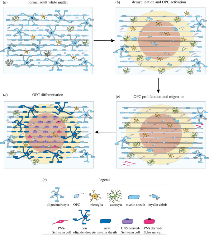
Figure 3.
Schematic of Schwann cell remyelination. (a) Under homeostatic conditions, astrocytes, microglia, oligodendrocyte progenitor cells (OPCs) and myelinating oligodendrocytes are dispersed throughout the normal adult white matter. SCs are absent from the CNS. (b) Following demyelination oligodendrocytes and myelin are lost. In some instances, astrocytes can also be damaged. Subsequently, OPCs in in the vicinity of the lesion area are activated. (c) Activated OPCs are recruited into the lesion area by the release of pro-migratory and mitogenic factors, and the demyelinated region is repopulated by new OPCs. At the same time, a small subset of peripheral SCs transgress into the CNS due to breaks in the glia limitans. (d) Typically, the lesion centre (red) contains the fewest number of surviving astrocytes, and in the absence of astrocytic inhibition, OPCs differentiate into SCs, which form a 1 : 1 association with the axon, resulting in a single myelin sheath. Conversely, oligodendrocyte differentiation and remyelination predominates at the lesion border (yellow), where astrocytes are present. In a minority of cases, peripherally derived SCs migrate into the lesion site, where they make contact and myelinate exposed axons. Remyelination is mostly complete within 3 weeks after lesion.

