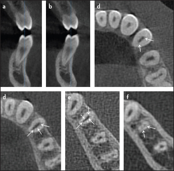
Figure 5.

Cross-section CBCT images of mandibular left first premolar showing one root with two canals (a, b). Axial CBCT image at the cervical third showing 2 canals (c), the middle third shows 3 canals (d), close to the apical third showing 4 canals (e) and 2 canals at the apex (f). (Arrows points to the canals). Canal configuration is 2-3-4-2
