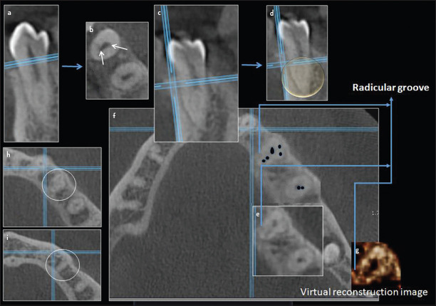
Figure 6.
Cross-section and axial CBCT images of mandibular right first premolar showing one root with two separated canals at the middle-third (a-d). Axial CBCT image at the middle third level (d) showing 5 canals (e) that was traced with black dots (f) and shown with virtual reconstruction image (g). More towered the apical third 4 canals can be seen (h) and 2 canals at the apex (i). See the radicular groove located at the mesio-lingual area (f). (Arrows points to the canals). Canal configuration is 2-5-4-2

