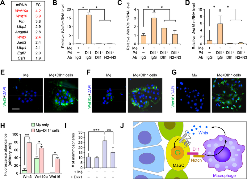
Fig. 8. Dll1 mediated crosstalk between MaSCs and macrophages promote Wnt ligand expression in macrophages to support MaSC activity.
(A) Fold-change in gene expression of the most differentially expressed genes encoding secreted factors or extracellular proteins between WT and Dll1cKO macrophage populations from mammary glands. (B-D) qRT-PCR analyses of expression of Wnt3, Wnt10a and Wnt16 in F4/80+ cells after co-culture with P4-Dll1+ cells with and without blocking antibody against Dll1, Notch2 and Notch3 antibodies. n = 3 samples, each with qRT-PCR in technical duplicate, and data are presented as the mean ± SD. (E-G) Representative IF images of co-culture cells (macrophages cultured for 3 days followed by addition of P4-Dll1+ cells for 5h) stained with Wnt3, Wnt10a and Wnt16 antibodies. Co-culture was washed extensively to remove P4-Dll1+ overlay cells from macrophages in short co-culture system. (H) Quantification of Wnt3, Wnt10a and Wnt16 immunofluorescence intensity in indicated groups from (E-G). Control was macrophage cultured alone without P4-Dll1+ cells. (I) Mammosphere assay of WT P4 cells with and without co-culture with macrophages along with Wnt inhibitor, Dkk1, n = 4 samples. For macrophage isolation, combination of F4/80 and CD140 antibodies were used. ***p < 0.001 and **p < 0.01 by Student’s t-test in (B-D) and (I). Mann Whitney U test was used in (H). Size bar, 10 μm in (E), (F) and (G). (J) Model showing crosstalk of Dll1+ MaSC enriched population with macrophages through Notch and Wnt signaling.

