Fig. 3.

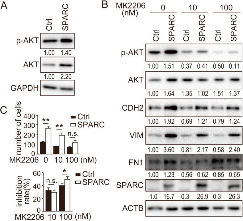
Induction of EMT and cell migration by SPARC is mediated by the AKT pathway. (A) Immunoblotting analysis of AKT and phosphorylated-AKT in SPARC-expressing Ishikawa cells. (B) Immunoblotting analysis of SPARC-expressing Ishikawa cells treated with indicated concentrations of the highly selective AKT inhibitor, MK2206. Antibodies against AKT, phosphorylated-AKT (p-AKT, Ser473), CDH2, VIM, FN1 and SPARC were used for immunoblotting. (A, B) Intensity of the bands was quantified using Image J. Values of the protein-of-interest were corrected using the intensity of GAPDH and ACTB bands, respectively. (C) In vitro cell migration of SPARC-expressing Ishikawa cells treated with indicated concentrations of MK2206. Numbers of migrated cells are shown in the top graph. Inhibition ratio of migrated cells compared with numbers of migrated cells at 0 nM is shown in the bottom graph. Ctrl, control; n.s., not significant; *, P < 0.05; **, P < 0.01. Full-length blot images are presented in Supplementary Fig. 4 and 5 (A, B). The experiments were independently repeated three times and representative data were shown
