Fig. 6.
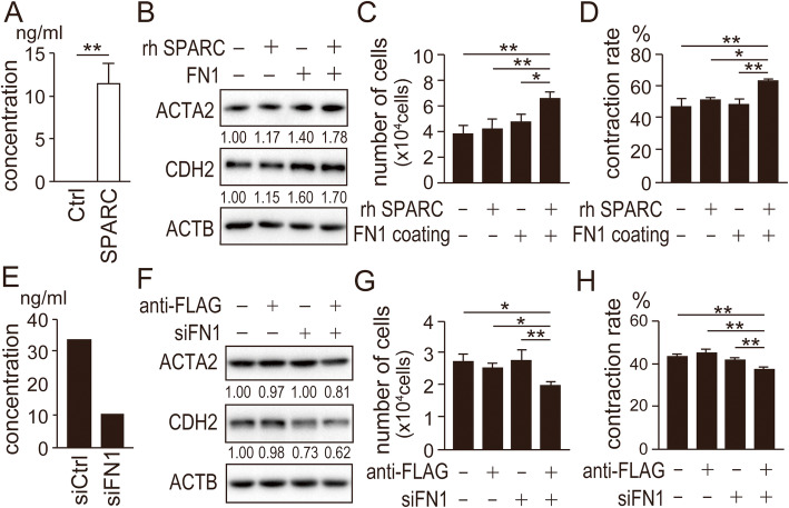
FN1 secreted from SPARC-expressing Ishikawa cells activates normal fibroblasts. (A) Amount of FN1 secreted from SPARC-expressing Ishikawa cells was measured by ELISA. (B) Protein levels of ACTA2 and CDH2 in NF cultured on FN1-coated dishes in the presence of recombinant SPARC were analyzed using immunoblotting. (C) NF were also analyzed for cell proliferation on day 6 in the same culture condition as (B). (D) NF cultured in the same condition as (B) were moved to collagen gel for in vitro contraction assays. (E) Successful knockdown of FN1 by siRNA (siFN1) was confirmed using ELISA. (F) Protein levels of ACTA2 and CDH2 in NF cultured in conditioned media from SPARC-expressing Ishikawa cells with siFN1 or immunodepleted of SPARC were analyzed using immunoblotting. NF were also analyzed for cell proliferation on day 6 (G) and in vitro contraction (H) in the conditioned media. (B, F) Intensity of the bands was quantified using Image J. Values of the protein-of-interest were corrected using the intensity of ACTB bands. Ctrl, control; rh SPARC, recombinant human SPARC; siCtrl, control siRNA; *, P < 0.05; **, P < 0.01. Full-length blot images are presented in Supplementary Fig. 10 and 11 (B, F). The experiments were independently repeated three times using identical fibroblasts and representative data were shown

