Fig. 1.
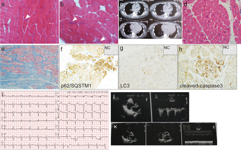
Histopathological findings of the skeletal muscle, cardiac tissue, and chest CT findings. Hematoxylin and eosin staining of the skeletal muscle showed scatter atrophic fibers (white arrow) and necrotizing myofibers (white arrowhead) (a × 200; b × 200). Enlarged heart was observed on the chest CT (c). Hematoxylin and eosin staining (d × 400) and Masson staining (e × 200) of the left ventricle revealed myofiber disarrangement, atrophy, and interstitial fibrosis with the absence of inflammatory infiltration. Immunohistochemistry analysis displayed p62/SQSTM1 was positive in some cardiomyocytes (f × 200). LC3 merely positively stained few cardiomyocytes (g × 200) and cleaved-caspase 3 mainly expressed on atrophic cardiac myofibers (h × 400) [inset negative control (NC) in f–h]. ECG demonstrated sinus rhythm, left anterior fascicular block and multifocal ventricular premature beat (i). Echo showed dilated left atrium and left ventricle (j in 2016 and k in 2019)

