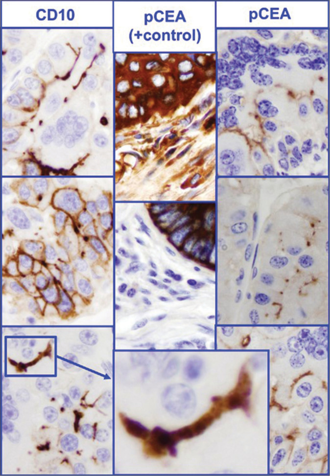
Figure 6:

Immunomorphology – bile canalicular pattern (Hepatocellular carcinoma). The immunostaining pattern highlights the bile canaliculi [by CD10 on left side or polyclonal CEA (pCEA) on right side] between different cells. The immunostaining should be seen as longitudinal sections with or without branching, Some may be seen as cross section and appear as dot between two cells. Nonspecific random immunostaining such as cytoplasmic immunostaining seen in the positive control in the center is interpreted as negative for bile canalicular pattern. The preferred positive control is liver tissue or hepatocellular carcinoma with reference to evaluation of bile canalicular immunostaining pattern.
