Figure 7:
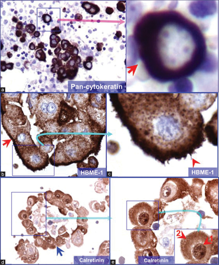
(a) Pancytokeratin immunoreactivity pattern (pleural fluid). Reactive mesothelial cells with cytoplasmic immunostaining (arrow in inset). Some reactive mesothelial cells may show a concentric immunostaining pattern around the nucleus better appreciated by adjusting fine focus. (b and c) HBME-1 immunoreactivity pattern (epithelioid mesothelioma, pleural fluid). Mesothelioma cells with membranous (arrow in a) and cytoplasmic immunostaining. Note the microvilli (arrowhead in b). (d and e) Calretinin immunoreactivity pattern (epithelioid mesothelioma, pleural fluid). Mesothelioma cells (arrow in a) show nuclear (arrowhead 1) immunoreactivity usually with cytoplasmic immunostaining (arrowhead 2) imparting the so-called “fried-egg” appearance (©vshidham reproduced from Ref #7).

