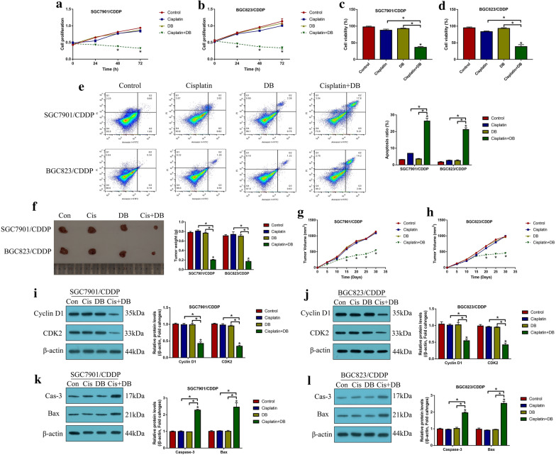
Fig. 1.
Low-dose DB sensitized CR-GC cells to cisplatin stimulation. The CR-GC cell lines (SGC7901/CDDP and BGC823/CDDP) were subjected to low-dose DB and high-dose cisplatin stimulation for 0 h, 24 h, 48 h and 72 h, respectively. a, b Cell proliferation and c, d viability were examined by CCK-8 assay and trypan blue staining assay. e Cell apoptosis was examined by Annexin V-FITC/PI double staining method. The xenograft tumor bearing mice models were established, and f tumor weight and g, h volume were examined, respectively. Western Blot analysis was conducted to determine the expression levels of i, j Cyclin D1 and CDK2, and k, l) cleaved Caspase-3 and Bax in CR-GC cells. (Note: “Con” indicated “Control”, “Cis” suggested “Cisplatin”, “DB” represented “Low-dose DB”, “Cis + DB” represented “Cisplatin plus low-dose DB stimulation”). Each experiment repeated at least three times. *P < 0.05

