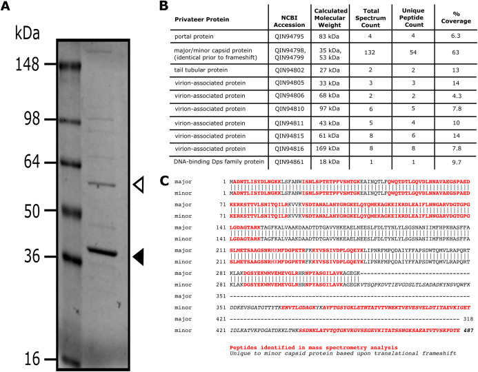
Figure 5. Analysis of Privateer virion proteins.
Proteins of phage Privateer identified by SDS-PAGE and mass spectrometry. (A) CsCl purified phage particles were separated on a 4–20% Tris-glycine SDS-PAGE gel. Molecular masses (in kiloDalton) of the protein ladder are displayed to the left of the gel. The white and black arrowheads indicate the expected location for minor and major capsid bands, respectively. (B) Table of mass spectrometry results for trypsin-digested Privateer proteins from whole phage particles. The total spectrum count is equal to the total number of total peptide spectral matches assigned to the protein, and the unique peptide count is equal to the number of peptide sequences exclusive to the protein. (C) Aligned sequences of the annotated major and minor capsid proteins. The highlighted regions indicate peptides identified via mass spectrometry.

