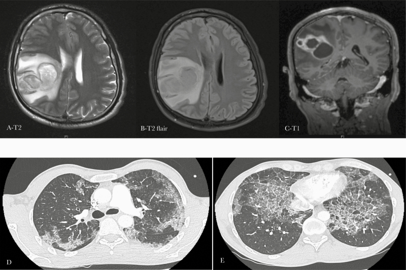
Figure 1.
Cerebral and thoracic imaging of a 40-year-old man with cerebral nocardiosis and pulmonary alveolar proteinosis. (A–C). Magnetic resonance imaging showing a voluminous cerebral parietal abscess. (D–E) Thoracic computed tomodensitometry scan showing diffuse and bilateral interstitial syndrome with thickening of the interlobular septa and a “crazy paving” aspect, which is typically found in pulmonary alveolar lipoproteinosis.

