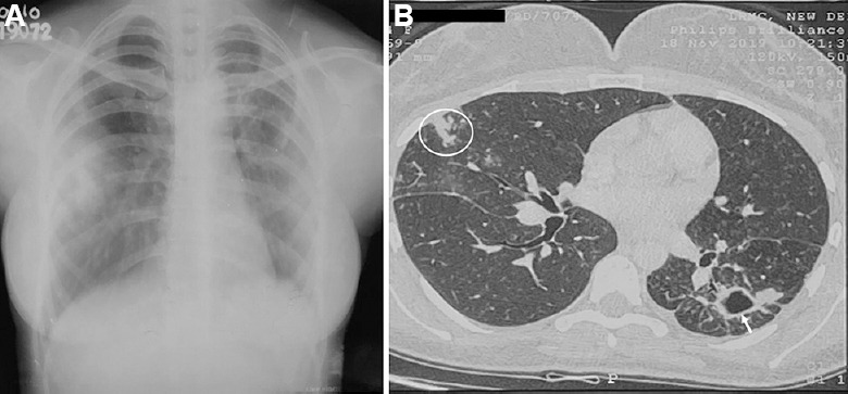
Fig. 3.

Chest radiograph in a 29 yr old female patient with Mycobacterium kansasii-pulmonary disease. (A) Chest X-ray reveals a cavitary lesion in the left lung. (B) Axial section in the high-resolution computed tomography scan demonstrates a cavity in the left lung (white arrow) and tree-in-bud appearance in the right lung (white circle).
