Figure 4.
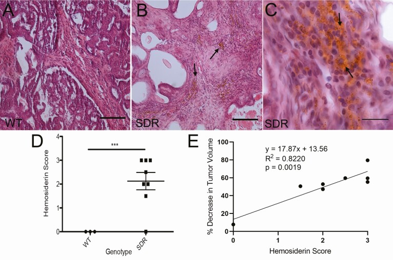
H&E staining of the mammary tumors in WT (A) and SDRs (B and C) treated with 1.25 mg/kg doxorubicin showed evidence of hemosiderin deposits (golden-brown stain) in SDRs indicated by arrows in B and C. (D) Blinded semiquantitative hemosiderin stain scoring indicates a complete lack of hemosiderin in WT rats (n = 3) with extensive evidence in SDRs (n = 8; ***P < 0.001; Student’s t-test). (E) Simple linear regression analysis shows a positive correlation between hemosiderin score and tumor volume decrease over the course of treatment in SDRs treated with 1.25 mg/kg doxorubicin. Scale bars in A and B = 100 µm. Scale bar in C = 20 µm.

