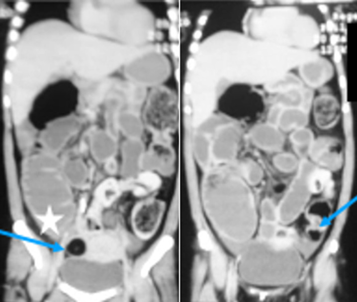
Figure 3.

coronal views of CT scan reveal dilated large intestine up to caecum (asterix) with no zone of transition, normal small bowel caliber, and no ascites or pneumoperitoneum. Incidentally, the patient was incidentally found to have bilateral adnexal dermoid cysts (blue arrows)
