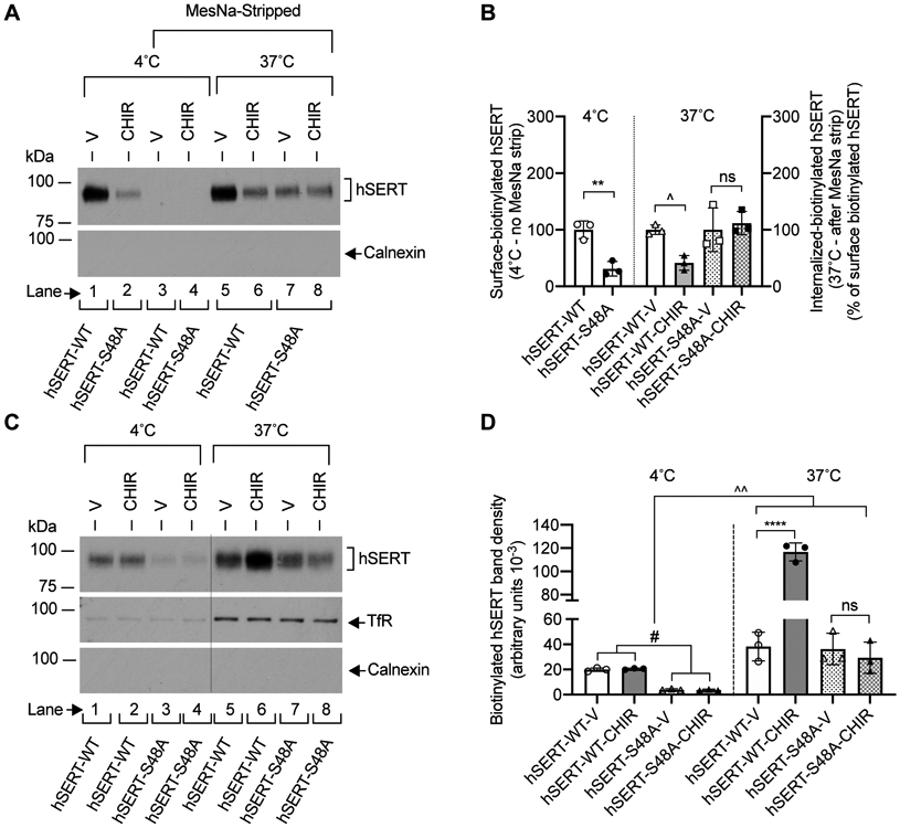
FIGURE 6. Effect of CHIR99021 on the internalization and plasma membrane insertion of hSERT-WT and hSERT-S48A in HEK-293 cells.

(A). Representative immunoblot of three independent experiments shows the surface-biotinylated hSERT before (lanes 1 and 2) and after MesNa treatment (lanes 3 and 4) (4°C), and internalized-biotinylated hSERT (37°C) after MesNa treatment (lanes 5, 6, 7 and 8). Intracellular marker calnexin from biotinylated fractions is also shown. (B). Quantified band densities (N=3 independent cell culture preparations, mean ± SD) of biotinylated hSERT bands (~94-98 kDa) reveal that CHIR99021 (2.5 μM) treatment for 30 min at 37°C significantly decreased hSERT-WT internalization, but had no effect on hSERT-S48A internalization (one-way ANOVA: F(5,21) = 8.581, P =0.0012). Bonferroni’s multiple comparison test: **P =0.0045, ^P =0.0138 compared between specified pairs. ns. non-significant (P =0.999) effect between indicated pairs. (C). HEK-293 cells expressing hSERT-WT or hSERT-S48A were incubated with biotinylating agent at 4°C (lanes 1, 2, 3 and 4) or 37°C (lanes 5, 6, 7 and 8) with vehicle or CHIR99021 (2.5 μM) for 30 min to biotinylate cell surface proteins. Calnexin and TfR levels were visualized following stripping the blots and reprobing with calnexin and TfR antibodies, and shown under hSERT immunoblot (C). Excised lanes 1, 2, 3 and 4 (4°C) from the same immunoblot were indicated with a vertical dashed line. A representative immunoblot of three independent experiments is shown. (D). Quantified biotinylated hSERT bands (N=3 independent cell culture preparations, mean ± SD) show that CHIR99021 treatment significantly enhanced hSERT-WT plasma membrane insertion, but had no effect on hSERT-S48A plasma membrane insertion (one-way ANOVA: F(7,16) = 61.85, P =0.0001). Bonferroni’s multiple comparison test: ****P <0.0001, ^^P =0.0014, #P =0.0115 compared between specified pairs. ns. non-significant (P =0.999) effect between indicated pairs. V: Vehicle, CHIR: CHIR99021, TfR: Transferrin Receptor.
