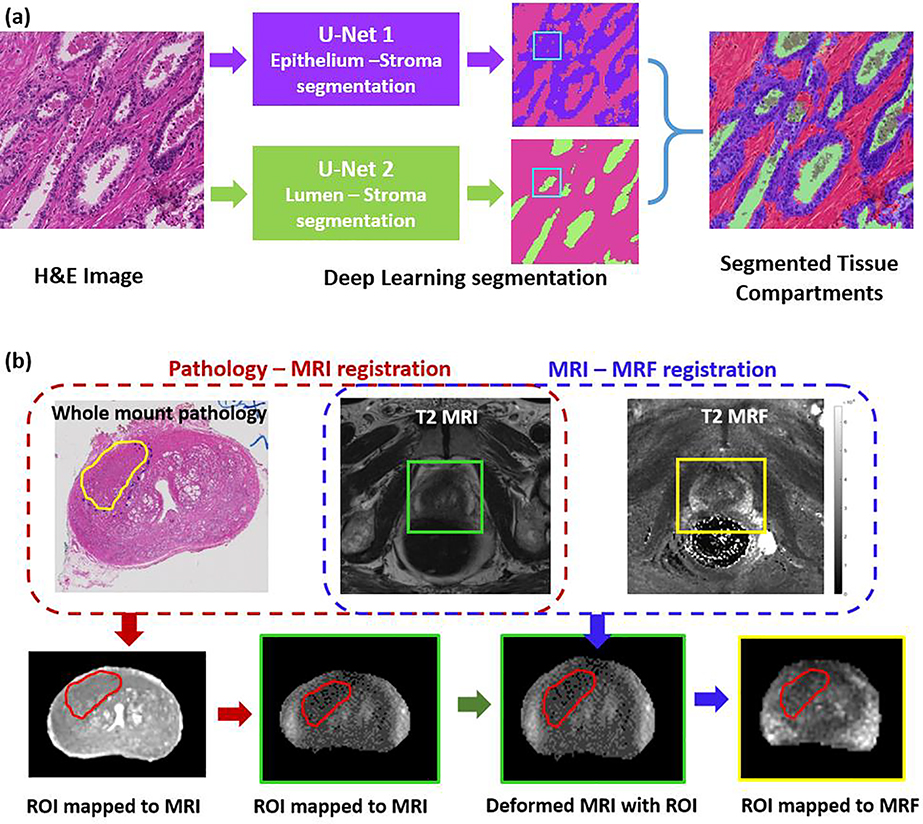
Figure 2:
(a) Tissue compartment segmentation on whole mount H&E stained digitized pathology using two U-net deep learning models. The lumen segmentation model has precedence when both the models are positive (an example is illustrated). (b) Schematic pipeline illustrating the workflow adopted to register digitized whole mounts with T2w images and T2 MRF. Prostate cancer and prostatitis ROIs delineated on pathology were mapped on to MRI and MRF following the registration.

