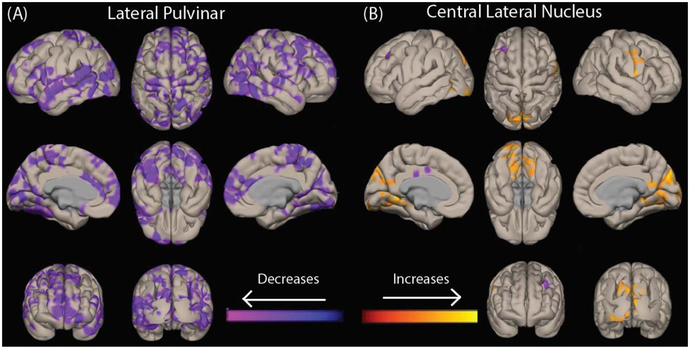
Fig. 2. Patients with temporal lobe epilepsy have different patterns of abnormal functional connectivity in key thalamic arousal nuclei.
Voxel-wise t-tests of functional connectivity seeded from bilateral lateral pulvinar (A) and central lateral nucleus (B) are shown in 40 patients vs 40 controls. Data for patients with right-sided epilepsy and their corresponding matched controls are flipped, so that changes ipsilateral to the epileptogenic side are shown on the left while contralateral changes are seen on the right side of the brain. Images are corrected for false discovery rate (FDR), and a cluster correction (p<0.05) is applied.

