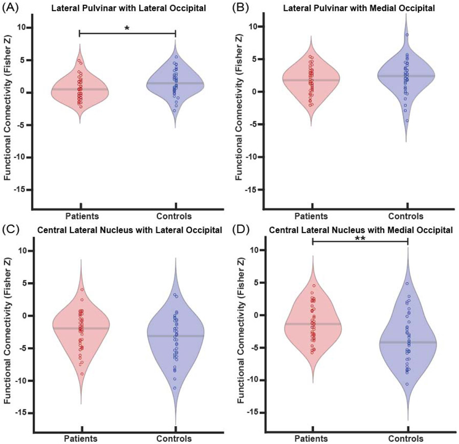
Fig. 3. Patients exhibit altered functional connectivity between thalamic arousal nuclei and regions of occipital lobe.
Patients demonstrate reduced connectivity between the lateral pulvinar and lateral occipital lobe compared to controls (A), but no differences in connectivity between the lateral pulvinar and medial occipital lobe (B). When examining central lateral nucleus connectivity, no connectivity differences to the lateral occipital lobe are seen (C), but connectivity to the medial occipital lobe is less negative in patients compared to controls (D). *p<0.5, **p<0.01, Bonferroni-Holm correction. N=40 patients with TLE vs 40 controls.

