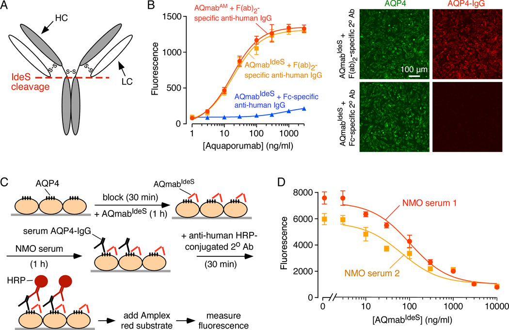
Figure 6.
AQmabAM competes with AQP4-IgG in NMO patient sera for binding to AQP4 on CHO-AQP4 cells. A. Schematic showing IdeS cleavage of IgG at its lower hinge region, producing F(ab’)2 and Fc fragments. B. (Left) Binding of AQmabAM (with or without IdeS treatment) to CHO-AQP4 cells. Fluorescence were detected by HRP-conjugated anti-human IgG, F(ab’)2 -specific, or Fc-specific, secondary antibody (mean ± S.E.M., n=3). (Right) Immunofluorescence showing binding of IdeS-cleaved AQmabAM (AQmabIdeS) to CHO-AQP4 cells using Cy3-conjugated goat anti-human IgG F(ab’)2 -specific, or Alexa 555-conjugated goat anti-human IgG Fc -specific secondary antibody. C. CHO-AQP4 cells were pre-incubated with AQmabIdeS for 1 h then incubated with NMO serum for 1 h, followed by HRP-conjugated anti-human IgG Fc-specific secondary antibody to detect serum AQP4-IgG binding. D. AQmabIdeS concentration-dependent displacement of AQP4-IgG in 1% NMO patient sera (mean ± S.E.M., n=3).

