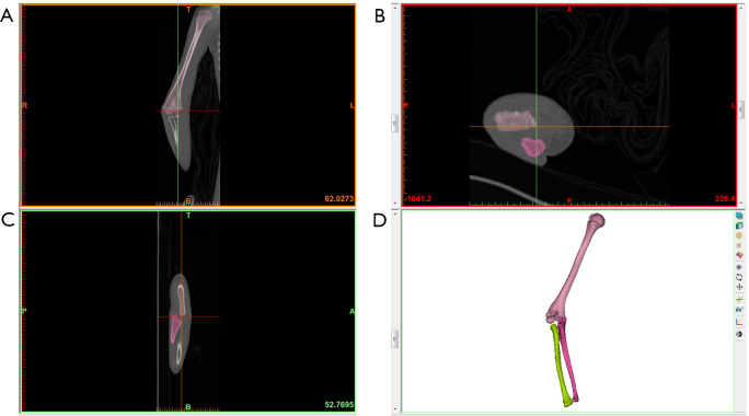
Figure 2.
Three-dimensional models of the affected bones were constructed in Mimics software. (A) CT shows the coronal section of the elbow; (B) CT shows the transverse section of the elbow; (C) CT shows the sagittal section of the elbow; (D) 3D reconstruction of elbow joints. CT, computed tomography.

