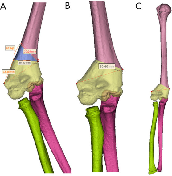
Figure 4.

The simulated closed-wedge osteotomy was done in Mimics software. (A) The first level of the medial hinge was closer to the joint immediately above the olecranon or coronary fossa; (B,C) a closing wedge osteotomy for angular correction was simulated, followed by correction on the osteotomy plane based on the deformity evaluation.
