
Figure 2.
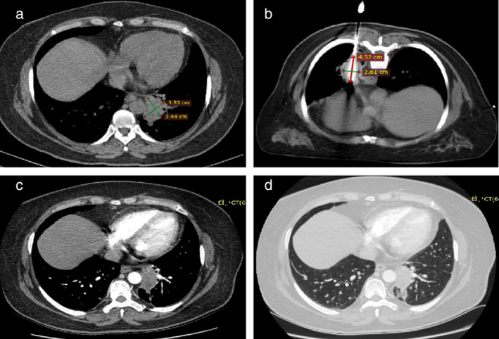
A 77‐year‐old woman with a 3.53 × 3.44 cm adenocarcinoma lesion received co‐ablation. (a) Initial computed tomographic (CT) scans before ablation show a lesion in the upper lobe of the left lung. (b) Co‐ablation was performed in the prone position using a probe. CT scan shows an iceball during co‐ablation. (c, d) Contrast CT scans one month after the procedure show no residual enhancement of the mass.