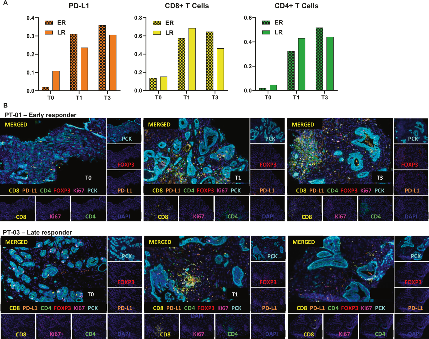
Fig. 3. Detection of human CD8 (yellow), CD4 (green), PCK (cian), FOXP3 (red), Ki67 (magenta) and PD-L1 (orange) in FFPE prostate cancer biopsies by IHC-IF.

a Quantification of PD-L1 (left), CD8+ (middle) and CD4+ (right) in multiplex images of early responders (ER) and late responders (LR) patients; Three patients per group. b Representative Multiplex IHC images of patients 1 and 3 (PT-01 and PT-03) at T0 (Diagnostic biopsy), T1 (Specimen collected after two cycles of nivolumab, and prior to HDR#1) and T3 (Specimen collected after 4 cycles of nivolumab and four weeks after HDR#1, but prior to HDR#2).
