Fig. 1.
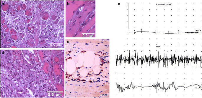
Diagnostic accuracy of muscle biopsy and electromyography in COVID-19-associated myopathy. Hematoxylin and eosin formalin-fixed and paraffin-embedded tissue sections from multiple biopsies of the left deltoid muscle and the left biceps brachial muscle. Panel a Scattered myofibers undergoing various stages of necrosis, with many atrophic muscular fibers, and nuclear centralization. Panel b An example of myophagocytosis. A macrophage (in blue) is phagocytizing a myofiber. Panel c immunostaining using CD68/PGM1+ antibodies demonstrates that the inflammatory infiltrate is mainly composed of macrophages. Scant lymphocytes B/CD20+ and T/CD3+ with a T-helper immunophenotype (CD4+) as well as CD123+ plasmacytoid dendritic cells were also found (data not shown). Panel d Other non-specific features are focal tissue sclerosis, atrophy, and variation in muscle fiber size. Panel e The electromyogram may be useful early in the diagnostic workup to confirm the presence of a myopathic pattern. Motor nerve conduction study from affected muscle (deltoid) shows compound muscle action potential (CMAP) amplitude reduced, with preserved distal latencies (top), reflecting muscle damage in the face of normal nerve function. Muscles tested by concentric needle (bottom) show short-duration, polyphasic low-amplitude motor unit potentials (MUP). Because each small motor unit is able to generate only a reduced amount of force compared with normal, with little muscle contraction, many MUPs are recruited. EMG traces were kindly supplied by Dr. Flavio Di Stasio, MD, and Dr. Giuliano Gentili, RNT

